Anatomy of Flowering Plants - Test Papers
CBSE TEST PAPER-01
CLASS - XI BIOLOGY
(Anatomy of Flowering Plants)
General Instruction:
- All questions are compulsory.
- Question No. 1 to 3 carry one marks each. Question No. 4 to 6 carry two marks each. Question No. 7 and 8 carry three marks each. Question No. 9 carry five marks.
1. Name two specialized kinds of parenchyma.
2. What is the function of companion cells in phloem?
3. Define meristem.
4. Why is cambium considered to be a lateral meristem?
5. Mention four characteristics of sunflower’s vascular bundles.
6. Differentiate between tracheids & vessels.
7. Explain the structure & function of collenchyma.
8. What are sieve elements? Explain their types & functions.
9. Describe the internal structure of a monocot root with the help of a labeled diagram.
CBSE TEST PAPER-01
CLASS - XI BIOLOGY (Anotomy of Flowering Plants)
[ANSWERS]
1. i) Aerenchyma ii) Chlorenchyma.
2. Companion cells help the sieve tube elements in translocation of food material, by maintaining their pressure gradient,
3. The specialised regions of plants where active cell divison continues are called meristems.
4. These meristems are present along the lateral sides of stem & roots therefore these are called lateral meristem. Cambium is considered as a lateral meristam as they are responsible for producing the secondary tissues on the plant and help in incerasing diameter og the plant part.
5. Sunflower is a dicot plant. Vascular bundles are conjoint, collateral, endarch, open and arranged in a ring surrounding pith (eustele), with xylem and phloem on the same radius, xylem being internal and phloem external. A strip of lateral meristem, cambium, is present between xylem and phloem.
Phloem lies outside of the vascular bundle and composed of sieve tubes, companion cells and phloem parenchyma (all are living) and few bast fibers. Companion cells are associated with sieve tubes.
Xylem lies toward the pith or center and composed of xylem vessels, tracheids, xylem fibres (wood fibres) and xylem parenchyma (wood parenchyma). The xylem is endarch (i.e. protoxylem lie towards the pith, while metaxylem lies towards periphery).
Cambium is a thin strip of two or three layered cells which are radically arranged. It lies between xylem and phloem.
6.
TRACHEIDS | VESSELS |
i) Found in all vascular plants | i) Found in angiosperms only |
ii) They are elongated or tube like daed cells with thick and lignified walls and tapering ends. | ii) Vessel is a long cylindrical tube-like structure made up of many dead cells called vessel members, each with lignified walls and a large central cavity. |
iii) Lumen is narrow | iii) Lumen is wider. |
iv) Tracheids have pointed ends. | iv) Vessel members are interconnected through perforations in their common walls. |
7. Collenchymas has polygonal cells & has unevenly thickened walls which are prominent at the corners. It is an example of simple tissue. Cells are more or less elongated with primary, non-lignified cell wall. The wall thickening is primary in nature & is composed of cellulose, hemicelluloses & pectin materials with high percentage of water. The thickening may be primarily at the corners or angles of the cells. They are found mostly in the hypodermis of herbaceous dicots in the form of homogenous layers or in the patches.
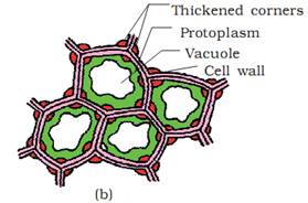
Function of Collenchyma:- The main function of this tissue is to give mechanical strength to the plant parts like young stem and petiole of the leaf. They also provide elasticity & support to the growing organs.
8. Sieve elements are the parts of phloem. They are meant for translocation & conduction of food material from leaves to different parts of the plant. Sieve elements are of two types:-
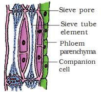
a) Sieve cells:- sieve cells are present in pteridophytes and gymnosperms. The cell wall is perforated. There are sieve plates throughout end walls & lateral walls.
b) Sieve tubes:- sieve tubes are present in angiosperms. Many sieve cells are connected to each-other to form a channel. There are sieve plates of the walls.
9. T.S. of monocot root shows the following tissues:-
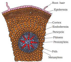
a) EPIDERMIS:- It is the outermost layer of root having no intercellular spaces stomata & cuticle. It bears unicellular root hairs.
b) CORTEX:- It is present beneath the epidermis. It consists of many layers of parenchymatous cells with large intercellular spaces.
c) ENDODERMIS:- It is the innermost layer of cortex. Its cells are barrel shaped with casparian strips on their antinunal walls. The passage cells are seen just opposite the protoxylem ends.
d) PERICYCLE:- It consists of single layer of thin walled parenchymatous cells.
e) VASCULAR BUNDLE:- The vascular bundles are radial, alternating xylem & phloem. The xylem & phloem bundles are always more than six. The xylem is exarch in condition. The central portion is occupied by large pith of parechyomatous cells. The conjuctive tissue is found between the xylem & phloem strand.